How Ptosis Repair Can Restore Eyelid Position and Improve Vision
September 5, 2026
Understanding Eyelid Ptosis and Its Impact on Vision
Ptosis repair is a surgical procedure that corrects a drooping upper eyelid by restoring the eyelid to its proper position, often improving both vision and appearance in the process.
Here is a quick overview of what you need to know:
- What it treats: A drooping upper eyelid caused by a weakened or stretched levator muscle
- Who it affects: Both children (congenital ptosis) and adults (acquired ptosis, most often age-related)
- Main surgical options: External levator surgery, Müller muscle conjunctival resection (MMCR), and frontalis suspension
- Non-surgical option: Prescription oxymetazoline eye drops for certain adults with mild acquired ptosis
- Why it matters: Left untreated, ptosis can impair vision, cause amblyopia in children, and lead to chronic neck strain from constant chin-up head tilting
A drooping eyelid is more than a cosmetic concern. It can quietly narrow your visual field, force your forehead muscles to work overtime, and in children, interfere with normal visual development entirely. You should not have to simply live with it.
I’m Dr. Shoib Myint, a board-certified Oculo Facial Plastic and Reconstructive Surgeon with over 20 years of experience and more than 15,000 surgeries performed, including a wide range of ptosis repair procedures for both functional and aesthetic outcomes. In this guide, I’ll walk you through everything you need to know, from understanding what causes ptosis to choosing the right surgical approach for your situation.
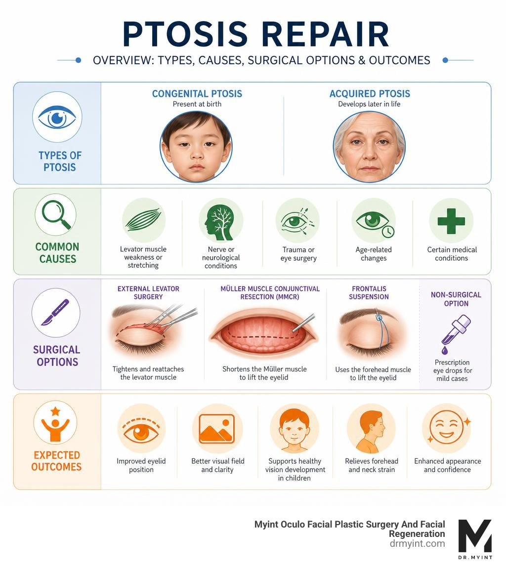
To understand how we correct a drooping eyelid, we must first look at why it happens. Medically known as blepharoptosis, ptosis occurs when the upper eyelid falls to an abnormally low position. This is primarily due to issues with the levator palpebrae superioris, the main muscle responsible for lifting the upper eyelid.
According to clinical resources on Ptosis(Archived) – StatPearls – NCBI Bookshelf, ptosis is broadly classified into two categories based on when it develops:
- Congenital Ptosis: This form is present at birth and is the most common type of ptosis overall. It typically stems from improper development of the levator muscle, which is often replaced by fibrous or fatty tissue. It seems to be more prevalent in males. If left untreated, between 20% and 70% of children with simple congenital ptosis will develop amblyopia, commonly known as lazy eye, because the drooping lid blocks light from entering the eye and disrupting normal visual development.
- Acquired Ptosis: This form develops later in life. Among acquired cases, aponeurotic ptosis is the most common type, usually presenting in late adulthood. It occurs when the tendon-like sheath of the levator muscle (the aponeurosis) stretches, thins, or pulls away from its normal attachment. This is often a natural part of the aging process, but it can also be accelerated by eye trauma, long-term contact lens wear, or previous eye surgeries, such as cataract extraction, where the medical instruments used to keep the eye open stretch the delicate eyelid tissues.
Other less common forms of acquired ptosis include neurogenic ptosis, caused by nerve damage, myogenic ptosis, linked to systemic muscle disorders like myasthenia gravis, and mechanical ptosis, where the eyelid is weighed down by a mass or excess skin.
When the eyelid droops significantly, it does more than alter your appearance. It actively encroaches on your superior visual field, making everyday tasks like reading, driving, or looking at a computer screen a constant struggle. You can learn more about how these changes impact your daily life by reading about whether Are Droopy Eyelids Impairing Your Vision? Consider an Eyelid Lift.
Surgical Techniques for Ptosis Repair and Comprehensive Facial Rejuvenation
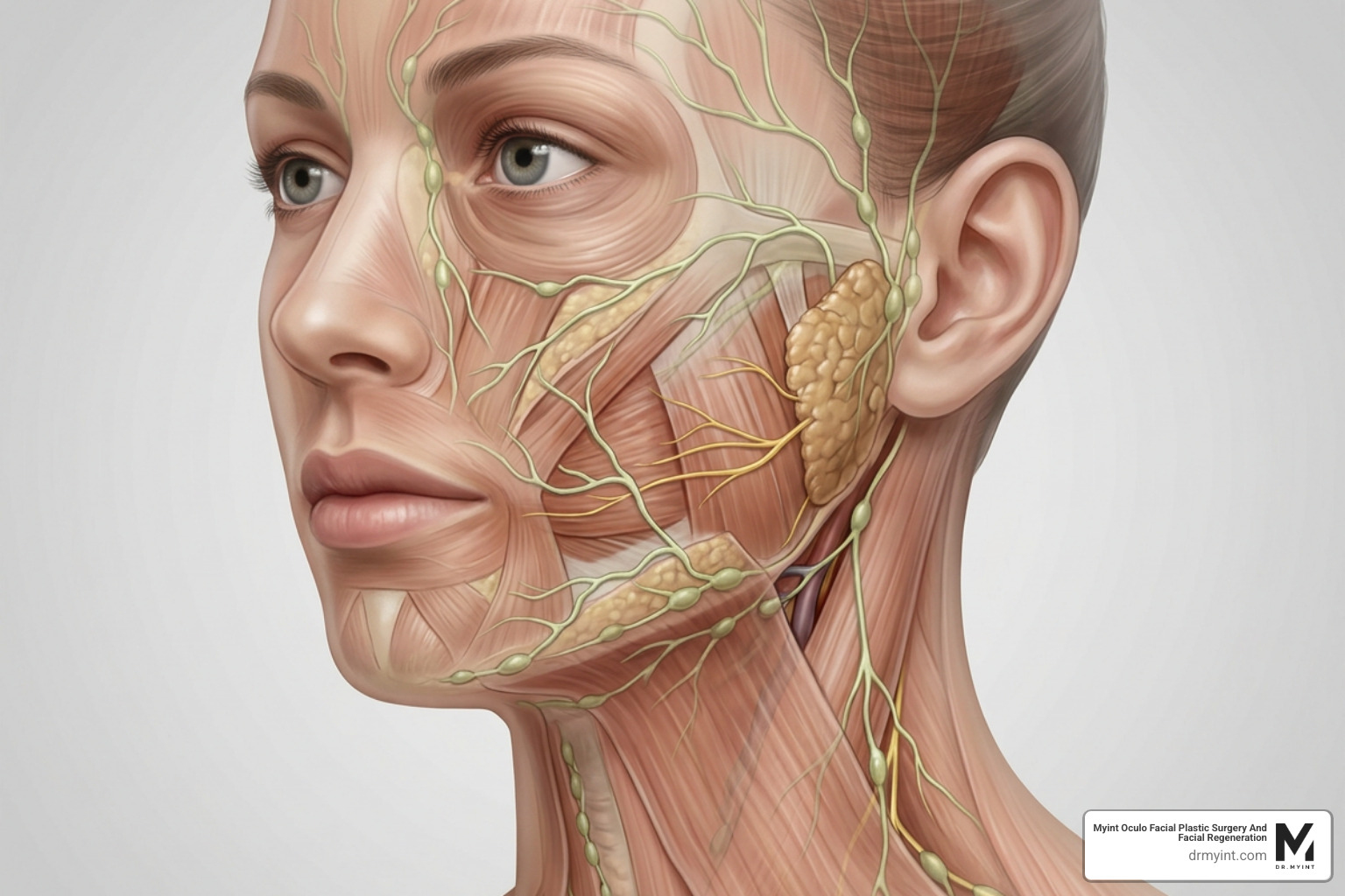
While correcting a drooping eyelid is essential for restoring your visual field, we rarely look at the eyes in isolation. True facial harmony requires evaluating how the eyelids interact with the rest of your facial anatomy.
As we age, the delicate structures around the eyes are not the only areas that lose structural support. The midface descends, jowls begin to form along the jawline, and the tissues of the neck lose their youthful firmness. Performing an isolated eyelid surgery on a face with significant structural sagging can sometimes create an unbalanced, unnatural appearance.
This is why we specialize in combining advanced oculoplastic procedures with comprehensive deep plane facelifts. By addressing both the delicate muscles of the eyelid and the deep structural layers of the face, we achieve a naturally refreshed, cohesive look that never appears pulled or operated on.
To understand the difference between adjusting the muscle that lifts the lid and simply removing excess skin, you can explore our guide on Ptosis Surgery vs. Blepharoplasty: How Are They Different?.
Anterior and Posterior Techniques for Ptosis Repair
When it comes to the actual ptosis repair procedure, surgeons have two primary pathways to access and tighten the levator muscle: the anterior (from the front) and the posterior (from the back) approaches.
- External Levator Surgery (ELS): This is an anterior approach. The surgeon makes an incision within the natural crease of the upper eyelid. Through this incision, the levator aponeurosis is identified, advanced, and secured to the structural plate of the eyelid (the tarsal plate) with sutures. This approach is highly versatile, allowing the surgeon to make large adjustments and simultaneously remove any excess skin or fat if a blepharoplasty is being performed.
- Müller Muscle Conjunctival Resection (MMCR): This is a posterior, internal approach. The surgeon flips the eyelid inside out to access the muscles from underneath. A specific portion of the conjunctiva and Müller’s muscle (a secondary muscle that helps elevate the lid) is resected and sutured. Because the incision is entirely on the inside of the eyelid, there are no visible external scars. This technique is highly precise and is typically chosen for patients who show a positive response to a preoperative phenylephrine eye drop test.
Selecting the right primary approach is critical, but sometimes adjustments are necessary. A comprehensive clinical study published in Revision plan and results of anterior and posterior ptosis surgery | BMC Ophthalmology | Springer Nature Link highlighted that revision surgery was required in 108 patients (15%) out of 780 patients who underwent MMCR or ELS for ptosis. The study demonstrated that if a primary surgery does not achieve perfect symmetry, switching techniques, such as moving from an internal posterior approach to an external anterior approach for the revision, is an exceptionally effective way to optimize eyelid height and contour.
For more details on what the surgical experience looks like, visit our dedicated page on Eyelid Ptosis Surgery.
Selecting the Right Approach for Ptosis Repair
The choice of surgical technique depends heavily on the severity of the ptosis and the measured strength of the levator muscle, known as levator function.
To measure levator function, we gently hold the eyebrow still to isolate the forehead muscles and measure how far the eyelid can move from extreme downward gaze to extreme upward gaze.
- Excellent to Good Function (9 mm or more): Patients with good muscle function are excellent candidates for either external levator advancement or internal MMCR.
- Fair to Poor Function (less than 5 mm): When the levator muscle is severely weakened or completely inactive, simply tightening it will not suffice. In these cases, we must utilize alternative techniques.
For patients with poor levator function, a Frontalis Suspension Procedure is often the gold standard. As detailed in the clinical guidelines on the Frontalis Suspension Procedure – EyeWiki , this technique uses a sling-type material to connect the eyelid margin directly to the frontalis (forehead) muscle. This allows the patient to lift their drooping eyelid simply by raising their eyebrows.
The choice of sling material is crucial. Autogenous fascia lata, harvested from the patient’s own leg, is considered the gold standard, with recurrent ptosis rates reported as low as 5%. Banked fascia lata is another option, carrying a recurrence rate of around 8%, while synthetic materials like silicone rods are highly popular because they avoid a second surgical site on the leg.
For patients with moderate to severe congenital ptosis who wish to avoid the potential forehead scarring and lid lag associated with traditional frontalis slings, advanced combined techniques offer an elegant alternative. A landmark study on a Modified Müller’s muscle conjunctival resection combined with levator plication in moderate to severe congenital ptosis with poor levator function – PMC demonstrated an overall success rate of 76.5% at a 6-month follow-up using this internal, combined approach, with 73.5% of cases achieving an excellent, natural eyelid contour without external incisions.
Integrating Deep Plane Facelifts for Total Facial Harmony
While correcting a drooping eyelid restores your visual field and brightens your gaze, true facial rejuvenation requires looking at the face as a whole. Eyelid aging does not happen in a vacuum. It is almost always accompanied by midface descent, the formation of jowls along the jawline, and a loss of definition in the neck.
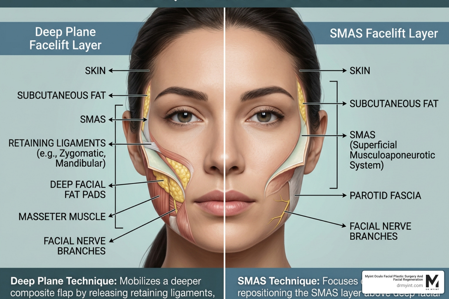
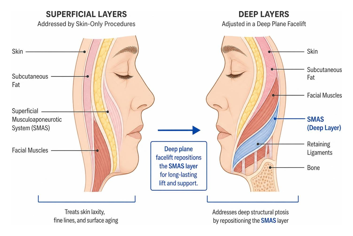
This is where the artistry of a deep plane facelift comes in. Unlike traditional facelifts that simply pull the skin tight, a deep plane facelift lifts the deeper muscle and tissue layers, specifically the Superficial Muscular Aponeurotic System (SMAS). By repositioning these deep structures vertically, we naturally lift the cheeks, smooth the nasolabial folds, eliminate jowls, and restore a sharp, youthful jawline.
When we integrate a deep plane facelift with a ptosis repair, we address facial aging at every level. The facelift provides the structural foundation and lifts the midface, while the eyelid surgery clears the visual axis and rejuvenates the eyes. This combined approach ensures that your upper face matches the youthful, lifted contours of your lower face and neck, creating a completely harmonious, natural result.
To explore how these procedures can be customized to your unique anatomy, learn more about our approach to Eyelid Lift Surgery.
The Role of Regenerative Medicine and Hyperbaric Oxygen in Eyelid and Facelift Healing
At Myint Oculo Facial Plastic Surgery and Facial Regeneration, our philosophy goes far beyond performing precise surgical cuts. We are deeply committed to utilizing the latest advancements in regenerative medicine to optimize and accelerate your healing process.
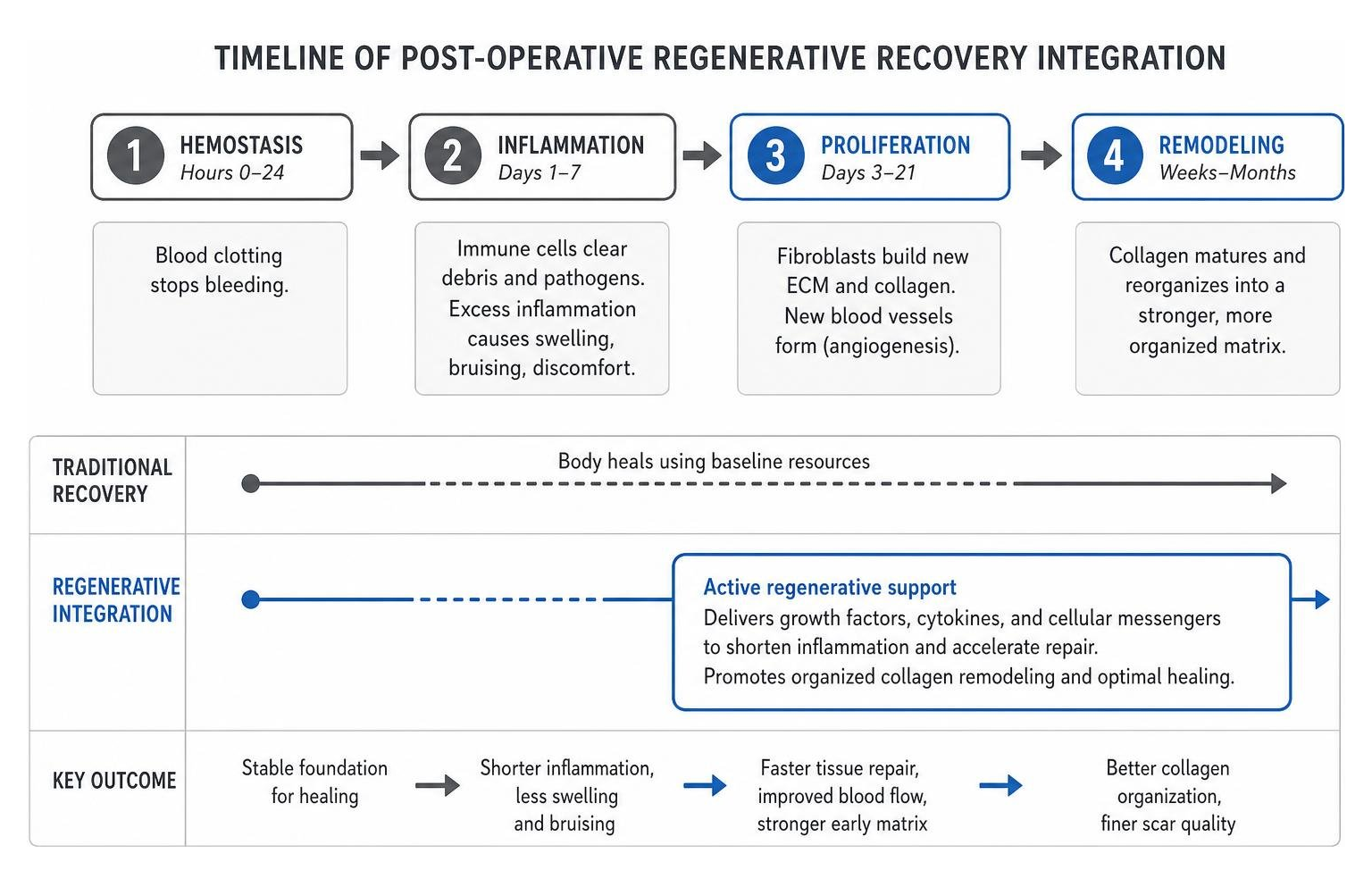
Major surgeries, particularly combined procedures like a deep plane facelift and ptosis repair, place a temporary demand on your body’s tissues. To heal beautifully, minimize swelling, and prevent noticeable scarring, your cells require a massive influx of oxygen and regenerative growth factors.
We utilize a multi-faceted regenerative protocol for our patients in Beverly Hills and Las Vegas:
- Platelet-Rich Plasma (PRP) and Cellular Therapies: During your procedure, we can apply concentrated growth factors derived from your own blood directly to the deep tissue layers. This stimulates rapid cellular repair, accelerates collagen synthesis, and promotes the healthy formation of new blood vessels.
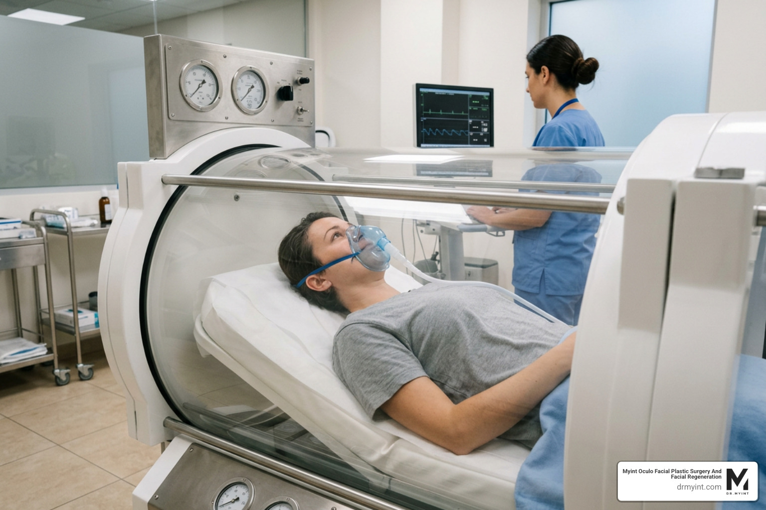
- Hyperbaric Oxygen Therapy (HBOT): We strongly recommend post-operative HBOT sessions. By breathing pure oxygen in a pressurized chamber, we increase the oxygen concentration in your blood plasma up to twenty times its normal level. This profound level of tissue oxygenation dramatically reduces swelling, speeds up wound healing, reduces the risk of infection, and helps scars fade into virtually invisible lines.
By combining these cutting-edge regenerative techniques with advanced surgical tissue handling, our patients experience significantly less downtime, allowing them to return to their busy lives feeling refreshed and looking completely natural.
What to Expect During Recovery from Ptosis Repair and Facial Surgery
Understanding your recovery timeline is key to a stress-free healing journey. While the thought of undergoing surgery on your face and eyelids can feel daunting, the actual recovery process is highly manageable when you follow a structured post-operative plan.
Most ptosis surgeries and deep plane facelifts are performed under local anesthesia with sedation, meaning you will be comfortable and relaxed during the procedure. Here is what you can generally expect during your recovery:
- The First Week: Swelling and mild bruising around the eyes and face are completely normal. This is a temporary phase of asymmetry as your tissues begin to heal. You will apply cool compresses to your eyes and keep your head elevated, even while sleeping, to minimize swelling. Post-operative follow-up appointments are typically scheduled within several days to one week after surgery to monitor healing and remove any external stitches.
- Weeks 2 to 3: Most of the swelling and bruising will resolve during this period. If you had a frontalis suspension, you may experience temporary lagophthalmos, which is an inability to fully close your eyelid during sleep. This is a common and expected part of the healing process that typically resolves within a few weeks as the muscles adapt. Daily use of lubricating eye drops and nighttime ointments is essential during this time to protect the surface of your eye.
- Activity Restrictions: To protect your delicate incisions and prevent complications, you must adhere to strict activity guidelines. This includes avoiding strenuous exercise, heavy lifting, and bending down for at least two weeks. Additionally, you must refrain from wearing eye makeup, drinking alcohol, or getting your incisions wet for the first couple of weeks. There is a strict restriction on swimming for at least four weeks post-surgery to prevent infection.
By practicing diligent hand hygiene, keeping your face out of direct sunlight, and attending all scheduled follow-up appointments, you will ensure a safe, smooth, and successful recovery.
Frequently Asked Questions about Ptosis Repair
To help you compare the primary surgical options we have discussed, here is a quick reference table:
| Surgical Approach | Incision Location | Best Candidate | Key Benefit |
|---|---|---|---|
| External Levator Surgery (ELS) | External eyelid crease | Moderate to severe ptosis with fair to good levator function | Highly adjustable; can be combined with cosmetic blepharoplasty |
| Müller Muscle Conjunctival Resection (MMCR) | Internal (under the eyelid) | Mild to moderate ptosis with good levator function | Completely scarless; highly predictable contour |
| Frontalis Suspension | Above the eyebrow and lid margin | Severe ptosis with poor to absent levator function | Restores lid elevation using the power of the forehead muscle |
Can a drooping eyelid be corrected without visible scars?
Yes. If you are a candidate for a posterior approach, such as a Müller muscle conjunctival resection (MMCR), the entire procedure is performed through a transconjunctival incision on the underside of the flipped eyelid. This internal approach leaves absolutely no visible external scars on the skin, allowing for completely discreet, scarless healing.
How does a deep plane facelift complement eyelid surgery?
A deep plane facelift addresses the structural foundations of the midface, jawline, and neck by lifting the deep SMAS layer. When combined with ptosis repair, it ensures complete facial harmony. Correcting a drooping eyelid refreshes your gaze, while the deep plane facelift restores youthful contours to the lower two-thirds of your face, preventing an unbalanced or “top-heavy” appearance.
What is the success rate of surgical eyelid correction?
Most surgeries to correct drooping eyelids are highly successful, yielding dramatic improvements in both visual fields and facial aesthetics. While a large-scale study noted a 15% revision rate among patients undergoing standard ELS or MMCR, these secondary adjustments are highly effective at fine-tuning eyelid symmetry. When performed by a dual-fellowship trained specialist, the long-term outcomes are exceptionally reliable and natural-looking.
Conclusion
A drooping eyelid should never stand in the way of your vision, your comfort, or your confidence. Whether you are dealing with a lifelong congenital condition or notice your eyelids gradually sagging with age, modern oculoplastic surgery offers precise, highly customized solutions to restore your natural eyelid position.
At Myint Oculo Facial Plastic Surgery and Facial Regeneration, we believe that the best results come from a deep understanding of both delicate eye anatomy and comprehensive facial rejuvenation. Dr. Myint’s rare dual fellowship training and over two decades of expertise in regenerative facial procedures allow us to offer unparalleled care, combining advanced surgical techniques with regenerative therapies to ensure a beautiful, natural, and rapid recovery.
If you are ready to restore your vision and achieve total facial harmony, we invite you to schedule a personalized consultation at our offices in Las Vegas, NV, or Beverly Hills, CA. Let us help you put your best face forward. Explore your options today by visiting our page on Eyelid Ptosis Surgery.